Serum Protein Electrophoresis
SPEP
- Is used to identify patients with multiple myeloma and other serum protein disorders.
- Reflects response to acute inflammation, malignancy, trauma, necrosis, infarction, burns, and chemical injury.
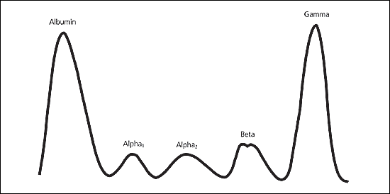
- A homogeneous spike-like peak in a focal region of the gamma-globulin zone indicates a monoclonal gammopathy.
Monoclonal gammopathies are associated with a clonal process that is malignant or potentially malignant, including- multiple myeloma
- Waldenström’s macroglobulinemia
- solitary plasmacytoma
- smoldering multiple myeloma
- monoclonal gammopathy of undetermined significance
- plasma cell leukemia
- heavy chain disease
- amyloidosis
- In contrast, polyclonal gammopathies may be caused by any reactive or inflammatory process.
- Normal Serum Protein Electrophoresis:
Positive electrodes (albumin), negative electrode (globulin)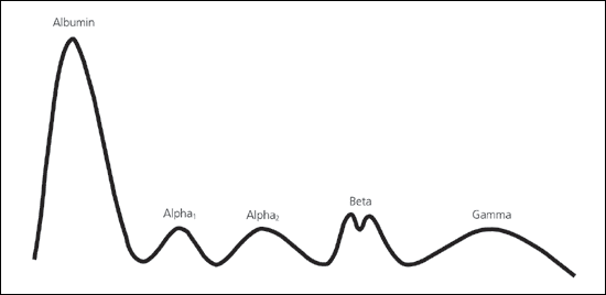
Albumin
Increased albumin:- Dehydration
- Chronic cachectic or wasting diseases
- Chronic infections
- Hemorrhage, burns, or protein-losing enteropathies
- Impaired liver function resulting from decreased synthesis of albumin
- Malnutrition
- Nephrotic syndrome
- Pregnancy
Alpha Fraction
It is comprised of alpha1-antitrypsin, thyroid-binding globulin, and transcortin. Malignancy and acute inflammation (resulting from acute-phase reactants) can increase the alpha1-protein band. A decreased alpha1-protein band may occur because of alpha1-antitrypsin deficiency or decreased production of the globulin as a result of liver disease. Ceruloplasmin, alpha2-macroglobulin, and haptoglobin contribute to the alpha2-protein band. The alpha2 component is increased as an acute-phase reactant.Increased alpha1 globulins
- Pregnancy
- Alpha1-antitrypsin deficiency
- Adrenal insufficiency
- Adrenocorticosteroid therapy
- Advanced diabetes mellitus
- Nephrotic syndrome
- Malnutrition
- Megaloblastic anemia
- Protein-losing enteropathies
- Severe liver disease
- Wilson’s disease
Beta Fraction
Beta1 is composed mostly of transferrin, and beta2 contains beta-lipoprotein. IgA, IgM, and sometimes IgG, along with complement proteins, also can be identified in the beta fraction.Increased beta1 or beta2 globulins
- Biliary cirrhosis
- Carcinoma (sometimes)
- Cushing’s disease
- Diabetes mellitus (some cases)
- Hypothyroidism
- Iron deficiency anemia
- Malignant hypertension
- Nephrosis
- Polyarteritis nodosa
- Obstructive jaundice
- Third-trimester pregnancy
- Protein malnutrition
Gamma Fraction
Majority of Immunoglobulins are found here, although they can be found throughout the electrophoretic spectrum.C-reactive protein (CRP) is located in the area between the beta and gamma components.
Increased gamma globulins
- Amyloidosis
- Chronic infections (granulomatous diseases)
- Chronic lymphocytic leukemia
- Cirrhosis
- Hodgkin’s disease
- Malignant lymphoma
- Multiple myeloma
- Rheumatoid and collagen diseases (connective tissue disorders)
- Waldenström’s macroglobulinemia
- Decreased gamma globulins
- Agammaglobulinemia
- Hypogammaglobulinemia
- Agammaglobulinemia
- Hypogammaglobulinemia
Indications
- Suspected multiple myeloma, Waldenström’s macroglobulinemia, primary amyloidosis, or related disorder.
- Unexplained peripheral neuropathy (not attributed to longstanding diabetes mellitus, toxin exposure, chemotherapy, etc.)
- New-onset anemia associated with renal failure or insufficiency and bone pain.
- Back pain in which multiple myeloma is suspected.
- Hypercalcemia attributed to possible malignancy (e.g., associated weight loss, fatigue, bone pain, abnormal bleeding)
- Rouleaux formations noted on peripheral blood smear.
- Renal insufficiency with associated serum protein elevation.
- Unexplained pathologic fracture or lytic lesion identified on radiograph.
- Bence Jones proteinuria
Interpretation
acute-reaction protein pattern: Response to acute inflammation, malignancy, trauma, necrosis, infarction, burns, and chemical injury is called “acute-reaction protein pattern” involves increases in fibrinogen, alpha1-antitrypsin, haptoglobin, ceruloplasmin, CRP, the C3 portion of complement, and alpha1 acid glycoprotein. Often, there are associated decreases in the albumin and transferrin levels.Monoclonal gammopathies
Disorders that are characterized by proliferation of a single clone of plasma cells that produce a homogeneous M protein. M protein is characterized by the presence of a sharp, well-defined band with a single heavy chain and a similar band with a kappa or lambda light chain. Once a monoclonal gammopathy is identified by serum protein electrophoresis, multiple myeloma must be differentiated from other causes of this type of gammopathy. Among these other causes are Waldenström’s macroglobulinemia, solitary plasmacytoma, smoldering multiple myeloma, monoclonal gammopathy of undetermined significance, plasma cell leukemia, heavy chain disease, and amyloidosis.- Multiple myeloma:
1. M protein appears as a narrow spike in the gamma, beta, or alpha2 regions.
2. M-protein level is usually greater than 3 g per dL.
3. Skeletal lesions (e.g., lytic lesions, diffuse osteopenia, vertebral compression fractures) are present in 80 percent of patients.
4. Diagnosis requires 10 to 15 percent plasma cell involvement on bone marrow biopsy.
5. Anemia, pancytopenia, hypercalcemia, and renal disease may be present.
Pattern in Multiple myeloma:
- Monoclonal gammopathy of undetermined significance
1. M-protein level is less than 3 g per dL.
2. There is less than 10 percent plasma cell involvement on bone marrow biopsy.
3. Affected patients have no M protein in their urine, no lytic bone lesions, no anemia, no hypercalcemia, and no renal disease. - Smoldering multiple myeloma:
1. M-protein level is greater than 3 g per dL.
2. There is greater than 10 percent plasma cell involvement on bone marrow biopsy. 3. Affected patients have no lytic bone lesions, no anemia, no hypercalcemia, and no renal disease. - Plasma cell leukemia:
1. Peripheral blood contains more than 20 percent plasma cells.
2. M-protein levels are low
3. Affected patients have few bone lesions and few hematologic disturbances.
4. This monoclonal gammopathy occurs in younger patients. - Solitary plasmacytoma:
Affected patients have only one tumor, with no other bone lesions and no urine or serum abnormalities. - Waldenström’s macroglobulinemia:
IgM M protein is present. Affected patients have hyperviscosity and hypercellular bone marrow with extensive infiltration by lymphoplasma cells. - Heavy chain disease:
The M protein has an incomplete heavy chain and no light chain.
Polyclonal Gammopathies
Monoclonal gammopathies are associated with a clonal process that is malignant or potentially malignant. In contrast, polyclonal gammopathies may be caused by any reactive or inflammatory process, and they usually are associated with nonmalignant conditions.Infections
- Viral infections: especially hepatitis, HIV infection, mononucleosis, varicella
- Focal or systemic bacterial infections, including endocarditis, osteomyelitis, and bacteremia, Tuberculosis
- Systemic lupus erythematosus
- Mixed connective tissue
- Temporal arteritis
- Rheumatoid arthritis
- Sarcoid
- Cirrhosis
- Ethanol abuse
- Autoimmune hepatitis
- Viral-induced hepatitis
- Primary biliary cirrhosis
- Primary sclerosing cholangitis
- Solid tumors
- Ovarian tumors
- Lung cancer
- Hepatocellular cancer
- Renal tumors
- Gastric tumors
- Lymphoma
- Leukemia
- Thalassemia
- Sickle cell anemia
- Gastrointestinal conditions, including ulcerative colitis and Crohn’s disease
- Pulmonary disorders, including bronchiectasis, cystic fibrosis, chronic bronchitis, and pneumonitis
- Endocrine diseases, including Graves’ disease and hashimoto’s thyroiditis
Abnormal SPEP in asymptomatic patient (Approach)
| Serum M-protein spike | ||||
|---|---|---|---|---|
| <1.5 gm/dl | 1.5 to 2.5 gm/dl | >2.5 gm/dl | ||
| SPEP yrly | Nephelometry | Metastatic Bone Survey, BM aspiration & biopsy, CT-Abdomen, beta-2 microglobulin level, CRP | ||
| Normal | Abnormal | Normal | Abnormal | |
| SPEP after 6mths | Ref. to Hematologist | SPEP after 3mths | Ref. to Hematologist | |